Ophioninae

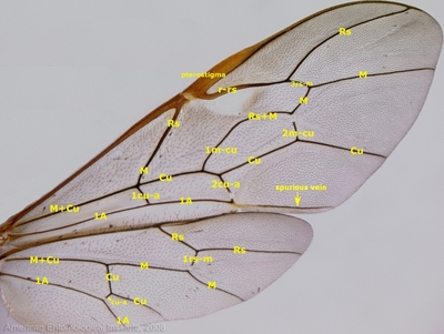
The fore wing of ophionines is distinguished by:

  1. a single 'intercubital' vein (presumably 3rs-m) that is apicad vein 2m-cu, and
  2. the presence of a spurious vein that extends apically from the apex of vein 1A.
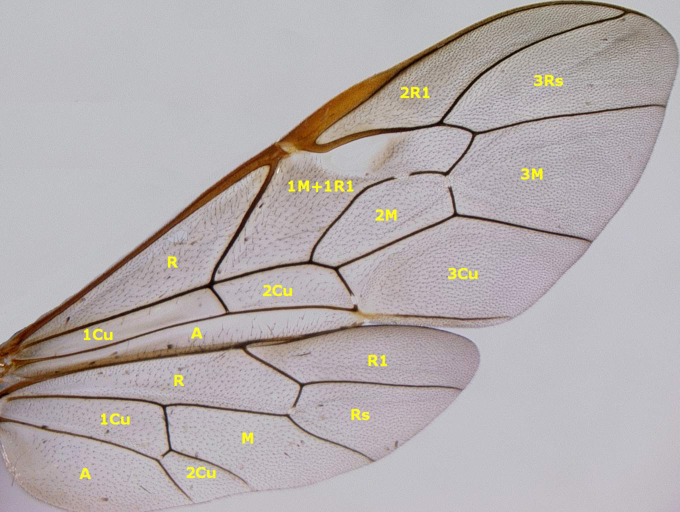
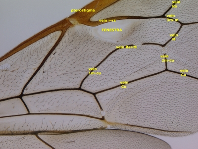
The veins and cells of Enicospilus texanus (Ashmead) are herein illustrated.


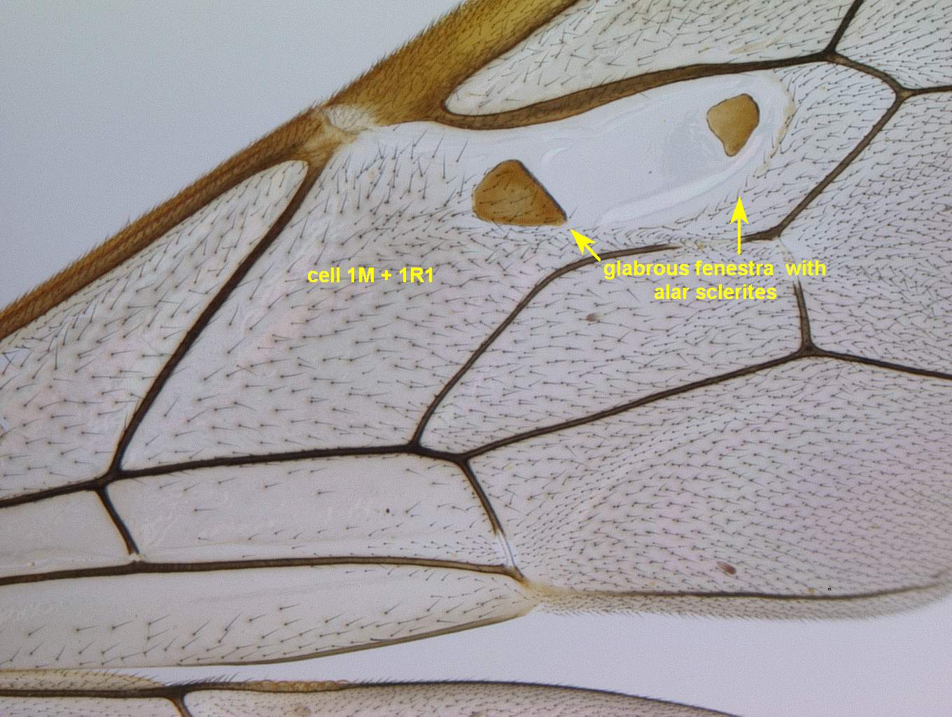
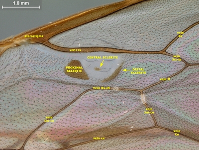
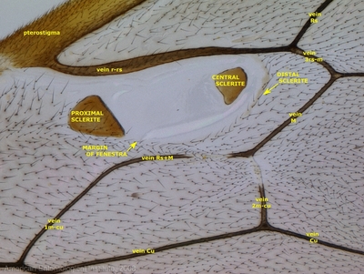
A number of genera possess a fenestra (a transparent glassy mark lacking setae) in cell 1M+1R1.
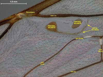
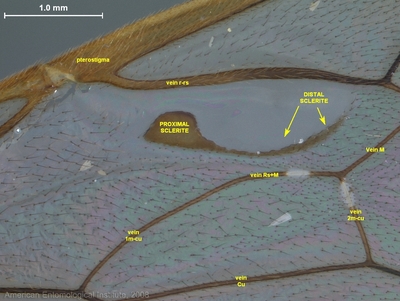
Many Enicospilus species have sclerotized inclusions (alar sclerites) within the fenestra.
The ground-plan condition consists of three alar sclerites (proximal, central, and distal) as shown in this specimen of Enicospilus flaviscutellatus.
There are modifications, however, as shown for Enicospilus purgatus (distal sclerite reduced to a thin crescent).
An undescribed Enicospilus from Florida (distal sclerite crescentic and attached to proximal sclerite)
Enicospilus guatemalensis (distal sclerite crescentic and attached to proximal sclerite, central sclerite absent).

Contents © 2002-present American Entomological Institute. All Rights Reserved.
Site credits and citation. Questions? E-mail: aei[at]aei.cfcoxmail.com